Application Sharing |Axia ChemiSEM in Hair Bundle
With the progress and development of the times, people's pursuit of beauty is no longer limited to the maintenance of facial skin. Whether the hand skin is delicate, whether the hair is healthy and shiny, and whether the hair volume is healthy can all affect a person's overall temperament. Hair is mainly composed of the epidermal layer, cortical layer, medullary layer, etc. The epidermal layer is mainly composed of many small scales overlapping, which is known as hair scales. It can protect our hair from external damage. Due to the limited resolution of the human eye, we cannot observe micrometer scale hair with the naked eye. With the help of the Axia ChemiSEM floor standing tungsten filament scanning electron microscope, we can observe the morphology and composition of hair scale, analyze its health level, and assist us in purposefully caring for our hair from the outside to the inside.
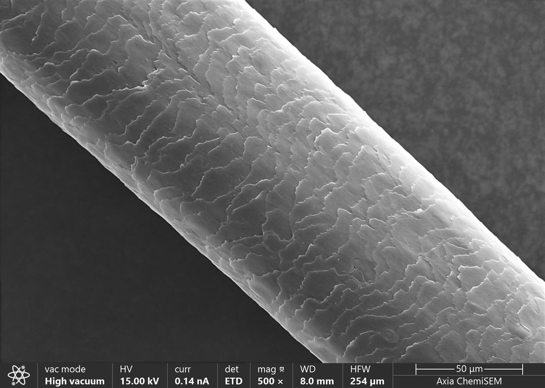
The continuous opening of hair scales can easily lead to the loss of nutrients from hair. The process of dyeing and perming can both lead to the shedding of hair scales, increased porosity, keratin degeneration, and nutrient loss. By using scanning electron microscopy to observe the morphology of hair scales on the surface of hair, one can observe its health level. We took the hair of three individuals as samples for analysis and comparison, named A, B, and C, to observe the impact of external factors on hair scales.
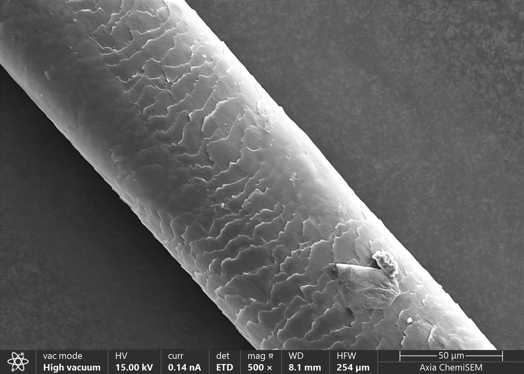
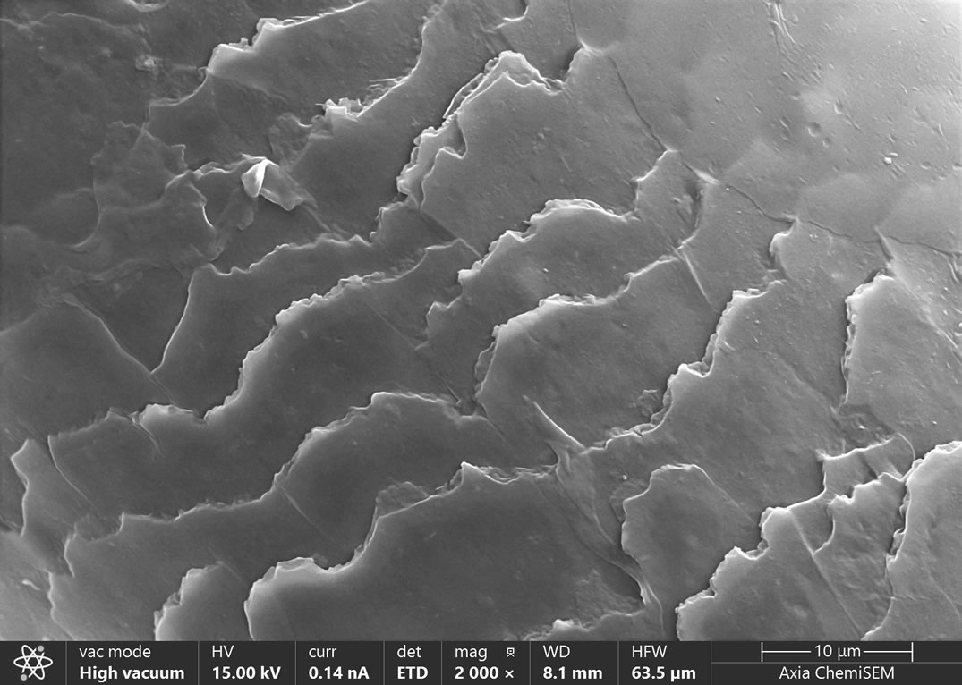
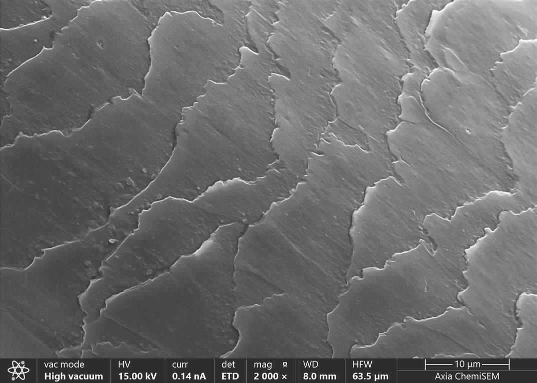
Firstly, sample A is hair that has been permed and sprayed with hair gel. The analysis results are shown in the following figure. Through electron microscopy analysis, we can observe that there is a certain degree of missing and uneven size of the hair scales on the surface of sample A, which may be caused by perm. At low magnification, it was observed that the scales on the entire hair thread were covered to varying degrees. Upon magnification, it was found that there were many pores on the surface of the cover, suggesting that it was caused by spraying hair glue. After spraying hair gel, the hair gel will cover the hair scales on the surface of the hair, making the hair scales closed and less prone to disorder, thus better shaping the hair.
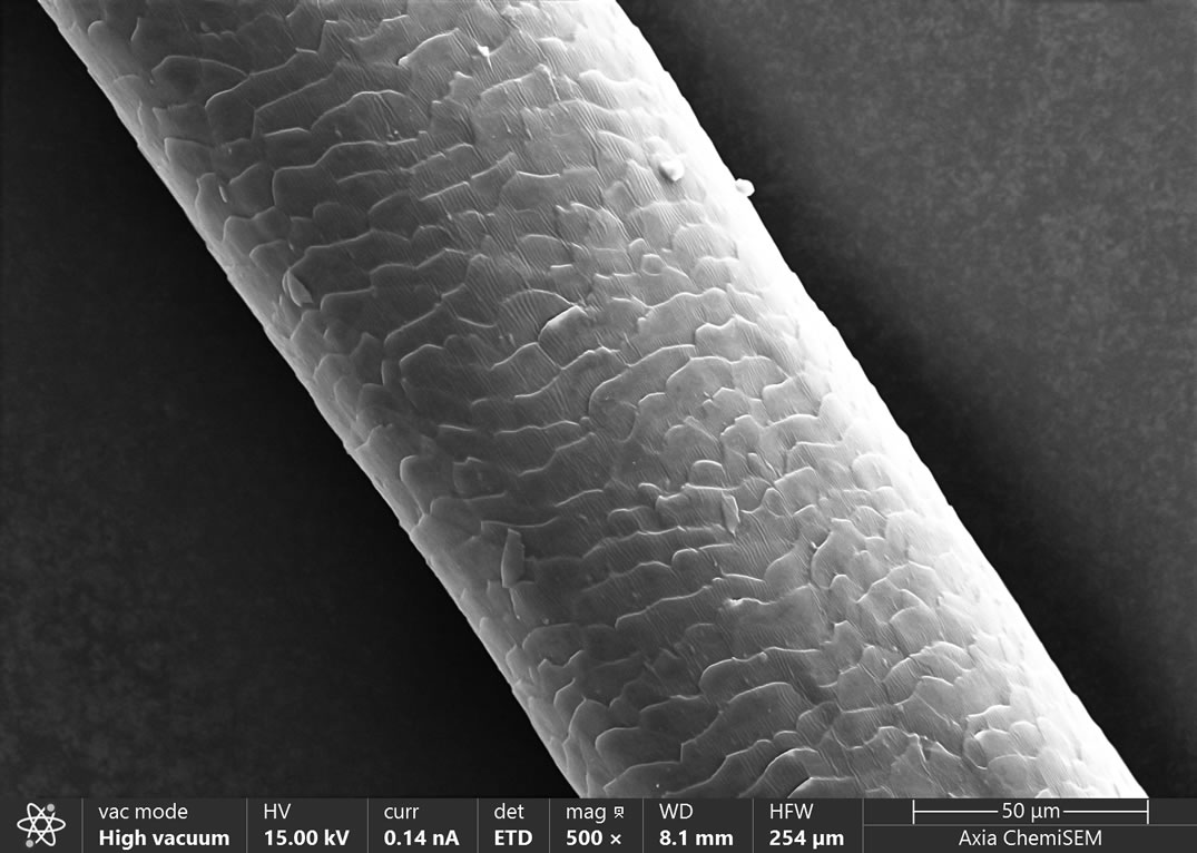
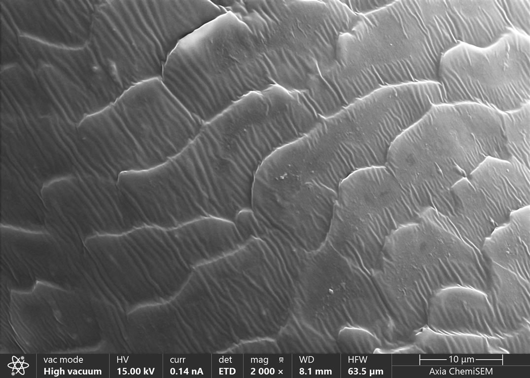
Secondly, we selected sample B treated with hair dye and conducted microscopic analysis on it. The analysis results are shown in the following figure. We observed through the images obtained by the Axia ChemiSEM tungsten filament scanning electron microscope that the entire hair's scales were tightly closed at low magnification. After magnification, we observed that the hair surface scales were covered with a layer of thin-film like substance, presenting a regular water ripple shape, and the scales were more closely attached to the hair shaft. Under normal circumstances, the hair scales in the hair bundle should be in an open state, and the hair scales treated with hair dye should be neatly closed together, greatly improving the phenomenon of curling hair.
Sample C without any treatment was selected for analysis, and the results are shown in the following figure. Through electron microscopy analysis, we observed that the hair scales on the surface of the hair were neatly arranged on the surface, showing an open state. Compared to A and B, untreated hair may be more prone to curling and rashness.
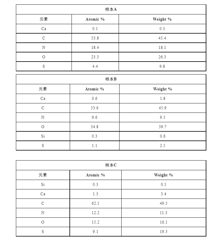
Axia ChemiSEM fully integrates various imaging modes of traditional SEM with real-time elemental quantitative analysis functions, allowing for rapid identification of various foreign objects over a larger area and obtaining real-time elemental analysis results with just one click. As shown in the following figure, the composition analysis results of the sample surface were obtained using Axia ChemiSEM, which includes both SEM and EDS data. The collection time is only a few minutes to obtain the composition information of surface foreign particles. We use the EDS element point analysis method to accurately and quantitatively analyze foreign elements. The Axia ChemiSEM user interface fully integrates all the functions of traditional EDS. When performing fixed-point element analysis on feature areas, we do not need to switch to other software to complete the analysis. Foreign objects on the sample surface can be detected at any time, greatly improving the analysis efficiency.
Using AxiaChemiSEM for elemental analysis of the sample, it was found that there was a significant difference in the elemental content between sample A and samples B and C. Sample A had a lower Ca content, while samples B and C had a higher Si content. This indicates that the sampler of sample A may lack calcium and need to consume more high calcium foods. B. The Ca content of C is normal, but the hair contains Si, indicating that the shampoo used contains silicone oil.
New generation Thermo Scientific ™️ Axia ™️ ChemiSEM intelligent tungsten filament scanning electron microscope can be used in low voltage and electron beam deceleration modes in conjunction with classic CBS probes (coaxial annular backscatter probes) to demonstrate the imaging performance of non-conductive samples under non gold plating conditions. Coupled with low vacuum mode, more realistic energy spectrum element analysis can be performed on non-conductive samples without coating. It can be used to observe the true morphology of various physically, chemically, and biologically treated samples, and then obtain morphology and composition information through scanning electron microscopy, providing customers with efficient solutions.